
This MSc thesis project is aimed at students in Artificial Intelligence, Computing Science, Data Science, or a related field who are interested in computer vision, biomedical data integration, and clinical decision-support systems.
Before organ transplantation, the immunological compatibility between a patient and a potential donor must be assessed carefully. An important aspect of this assessment is the detection of antibodies directed against Human Leukocyte Antigens (HLA), highly polymorphic cell-surface proteins that play a central role in antigen presentation and immune recognition. Pre-existing antibodies against donor HLA molecules can increase the risk of antibody-mediated organ rejection. Within HLA diagnostics, several complementary laboratory techniques are used to evaluate this compatibility.
Examples include:
Together, these tests provide important information about the immunological profile of a patient and the potential risk of incompatibility with a donor. At present, however, these data are generated in different laboratory workflows and are often stored in separate software systems. This means that laboratory specialists need to manually combine and interpret information from multiple sources to obtain a complete picture of the patient's immunological status.
At the same time, part of the CDC workflow is still assessed manually using fluorescence microscopy. This project therefore investigates how Artificial Intelligence, computer vision, automation, and data integration can be used to improve both the generation and interpretation of immunological laboratory data. The project consists of two closely connected parts.
In a CDC assay, patient serum is incubated with lymphocytes and complement. If relevant antibodies are present, complement activation may cause cell death. Fluorescent dyes are used to distinguish:
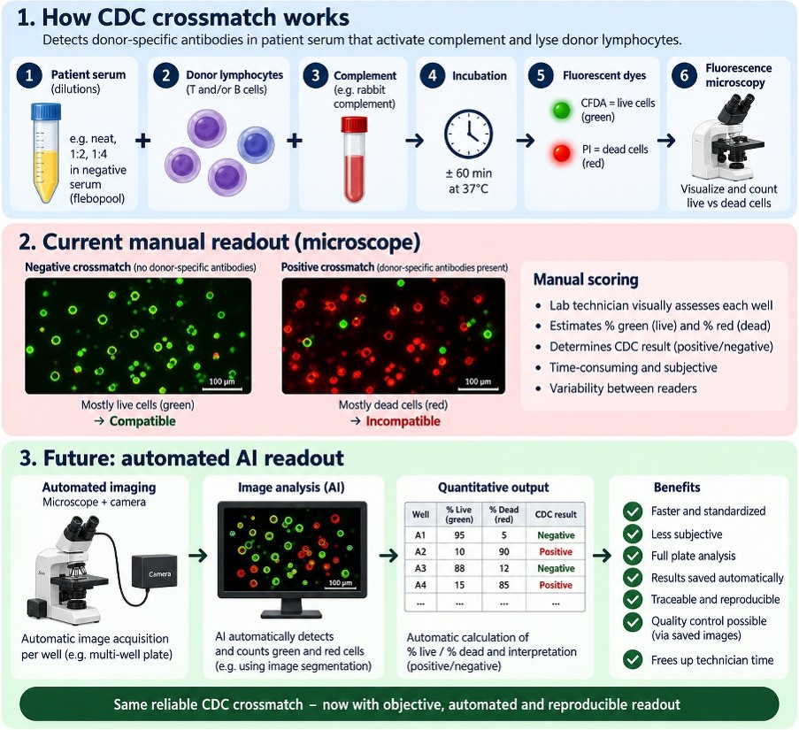
Currently, a laboratory technician evaluates the wells manually under a fluorescence microscope and visually estimates the proportion of living and dead cells. Although this method is well established, manual evaluation is time-consuming and introduces reader-dependent variability.
The first aim of the project is to develop a prototype for automated imaging and analysis of CDC assays. The envisioned workflow is:
CDC well → microscope + camera → automatic image acquisition → AI image analysis → % live/dead cells → CDC score → stored image and result.
The student may investigate classical computer-vision methods, machine-learning approaches, deep learning, or a combination of these techniques.
CDC results represent only one part of the patient's immunological profile. Additional information is obtained from several laboratory techniques, including:
At present, these results are often located in different software applications and data formats. A laboratory specialist therefore has to move between multiple systems to obtain a complete overview.
The second aim of this project is to investigate how these heterogeneous sources of immunological data can be brought together into a single integrated patient overview. The envisioned concept is:
HLA typing + LMX + LSA + CDC → data integration → immunological patient overview
The system should provide a clear and intuitive representation of the available immunological information for an individual patient. Rather than replacing expert interpretation, the goal is to create a tool that allows laboratory specialists to see the relevant information in one place, identify relationships between different test results more easily, and support consistent interpretation.
We are looking for a motivated MSc student with experience or interest in one or more of the following areas:
Experience with technologies such as Python, OpenCV, scikit-image, PyTorch, TensorFlow, pandas, SQL, or web-based visualization frameworks is an advantage. Previous knowledge of HLA, immunology, or transplantation is not required. The necessary biological and laboratory background will be provided during the project.
This project provides the opportunity to work on a real clinical laboratory problem with direct practical relevance within the Transplantation Immunology group in RadboudUMC. You will work with:
If you are interested, send your CV to dr. Cresci-Anne Croes or to dr. ir. Gabriel Bucur.